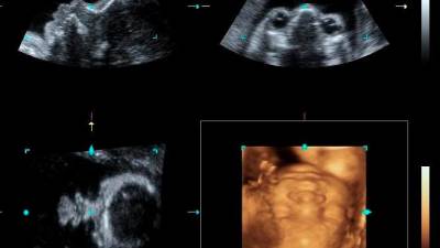
A detailed scan or an anomaly scan is an ultrasound examination done to look at the anatomical or physical structure of the baby. It can be a diagnostic test to check the physical structure or abnormalities in the baby.
It can also be used to screen for chromosomal abnormalities, but this will then require a diagnostic test such as amniocentesis. During the test, we look for soft markers that are not real abnormalities, but to show an increased risk of chromosomal abnormalities.
It is recommended to be done between 18 to 22 weeks of pregnancy. However, with better training and machines, most of the major abnormalities can be detected at 14 weeks of pregnancy but it is still recommended to repeat at 18 to 22 weeks of pregnancy especially in the heart where only about 50% of abnormalities can be detected in the best hands.
Here the whole structure of the baby that includes the head, brain, face chest, heart, stomach, kidneys, bladder, spine and all four limbs are checked to be anatomically normal.
This scan is best done by a maternal fetal medicine specialist. However, an obstetrician and gynaecologist that is trained and credentialled can also do these scans.
However, general practitioners are not trained to do these examinations. By law, they and the sonographers (not registered under the Private Healthcare and Facilities Act) are not allowed to perform these examinations as they are unable to provide the complete test.
These examinations are not only about the scan, but also about counselling. Ideally, the patient should be counselled on the implications of the test in this case the ultrasound examination. They should be counselled on the implications of a finding when the scan shows an abnormality.
Even when the scan does not show an abnormality, the patient needs to be informed that certain abnormalities may develop later or not detected due to the fetal circulation such as an atrial septal defect and a persistent ductus arteriosus, which is normal in a fetal circulation but not after birth. The scan also does not detect chromosomal abnormalities and rare genetic syndromes.
If there is an abnormality detected, the patient may require a referral to a neonatologist and a paediatric surgeon. Counselling can be done together by all of these professionals if the team approach is used.
The plan for the management of the baby can be executed before the baby is born. This ensures a smooth plan and execution of the treatment of the baby, in which care in the neonatal intensive care unit and the surgical management are done together.
If there is a query about the cause of the abnormalities being due to a chromosomal abnormality, then an amniocentesis with an appropriate laboratory test will be chosen for the best identification of the problem.
This could also shed on the risk of other problems in the baby that will be counselled to the patient and her family.
After that, a proper counselling with all the risks and possible treatment plans will be discussed with the patient and her family. The plan after agreement and consent can then be carried out for the best interest of the baby and family.
There are options available to patients and some of the treatment may even be initiated while the baby is still in the womb.
These treatments include medications, intrauterine blood transfusion for fetal anaemia, pleural tapping or chest tube for babies who have fluid in the lungs and a suprapubic catheter in those that cannot pass urine.
This article is contributed by consultant obstetrician & gynaecologist, maternal fetal medicine Datuk Dr H. Krishna Kumar.